
Bert Myers
I am a retired academic physician (Professor of Surgery at LSUMC) who has long had a serious interest in photography as an art medium. Beginning in 1951, I worked with Eugene Delcroix, a N.O. photographer who was a master of light and composition, and who specialized in soft focus images. In 1971, I studied with Ansel Adams in California at one of the last courses the artist himself taught. I have also been a pupil of Michael A. Smith of Ottsville, Pennsylvania-a master of large format photography-who in 1986 photographed New Orleans for the Historic collection. In 1999 I participated in a Santa Fe workshop on digital photography.
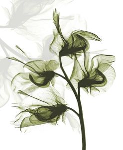
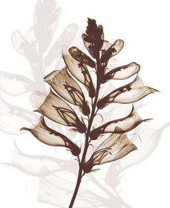
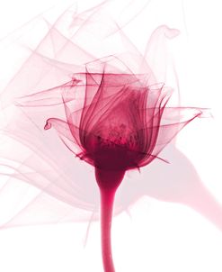
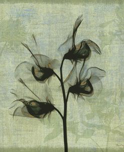
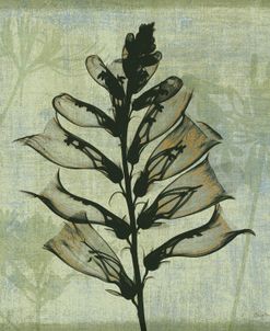
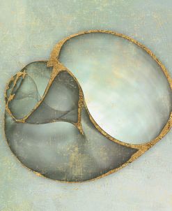
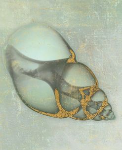
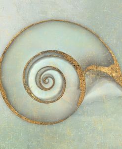
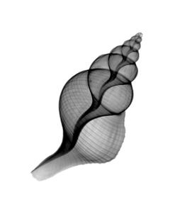
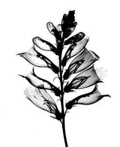
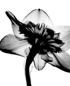
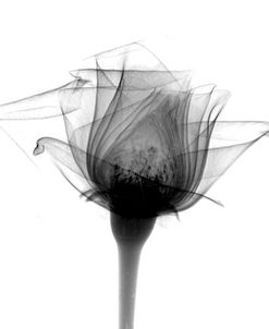
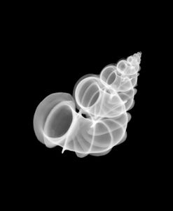
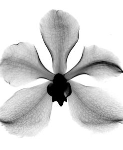
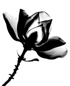
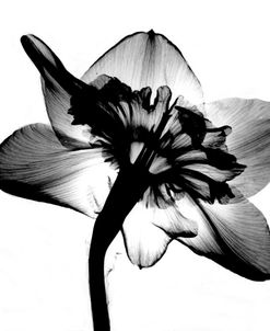
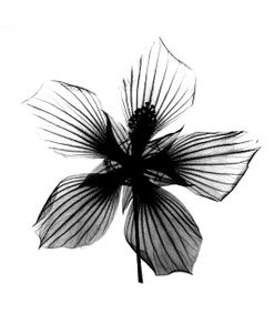
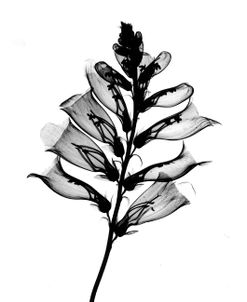
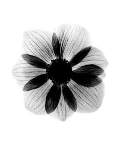
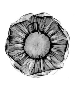
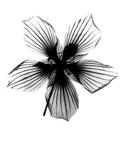
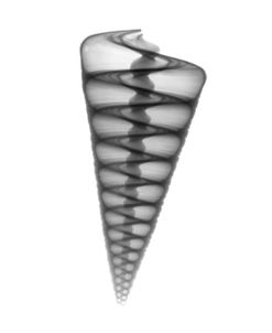
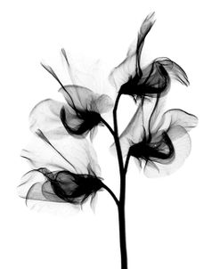
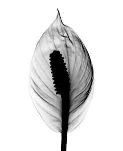
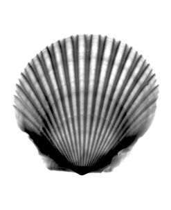
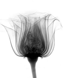
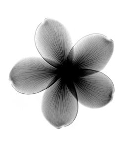
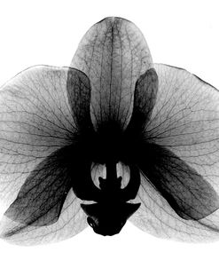
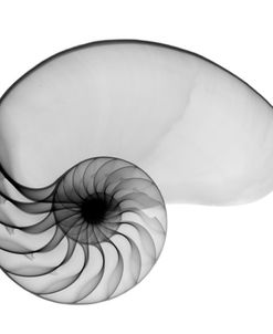
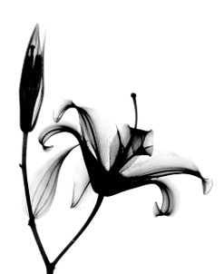
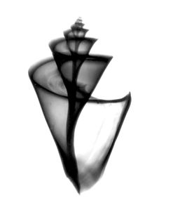
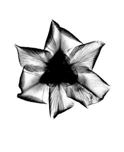
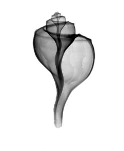
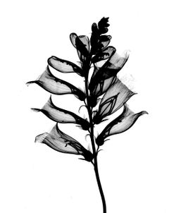
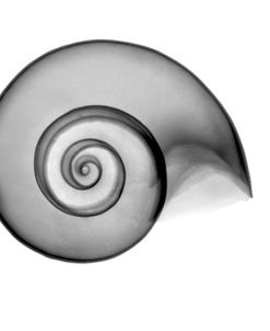
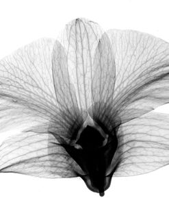
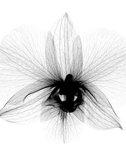
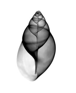
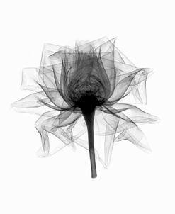
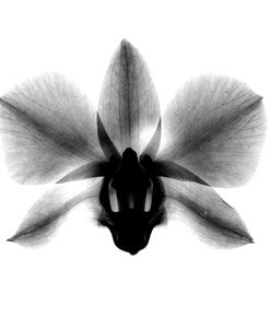
My first one man show was in 1976, at the Gallery Studio 8 in New Orleans. The images in that exposition were mostly scenics in the Adams tradition. In 1978, I began experimenting with using X ray as an art medium, developing the technique of taking radiographs of shells, flowers, and other objects and making B + W positive prints of them. Although the technique is not original, it is quite difficult and not widely done. I have written an article describing the technique, which was published in Applied Radiology, and articles about my methods have appeared in Medical World News, Art Center and the Free University in N.O., and for 9 years taught a course in advanced darkroom techniques in the Univ. College of Tulane University.
Since 1986 I have been using Cibachrome techniques to make radiographic images in color, including montages of straight photo images with xrays.
Beginning in 1985 I worked with high contrast internegatives and have a large collection of line derivations and solarizations, mainly of historic buildings.
From 1987-1997, I was interested in holography and took courses in the subject from Dr. T. Jeong at Lake Forest College in Illinois and Jeffrey Murray at the Holography Institute in Petaluma, California. I started the Holography research Laboratory at the Veterans Affairs Medical Center in N.O. and pursued the uses of holography in medicine, especially in education. I have developed techniques of making 3D images of bones, medical models, and human tissue which had been preserved by plastination, and had shows of my work in galleries in the USA, Scotland, and Hungary. My goal was to make a medical text illustrated with holograms. Unfortunately the field of educational and art holography did not progress. The extreme difficulty and cost of making and reproducing the images and the need for special lighting to view them impeded growth. In 1997, AGFA, the main producer of high quality holographic films and plates discontinued production, and I decided to abandon holography and concentrate my work in conventional photography, especially using digital technology to make color prints of Xray images.